
We remember things, words, and experiences from the past with the help of our memory. Episodic memory, damaged in some dementias and neuropathologies, stores all we have recently done and is an essential part of our identity as individuals.
The risk of forgetting or not remembering increases with age and is one of the common manifestations of Alzheimer’s and other dementias. Storing information and forming episodic memories involves different parts of the brain and the dorsal prefrontal area has a principal role. The dorsal left prefrontal cortex, a large and complex brain region, is relevant for cognitive performance and is involved in working memory organization.
In a recently published study in Plos Biology, Mirceva van der Plas and Simon Hanslmayr from Glasgow University, UK, and the Strungmann Institute (ESI) for Neuroscience in Germany, and colleagues, showed after stimulating the brain in the left prefrontal cortex the episodic memory improved (Figure 1).
In their experiment, researchers compared the ability to remember words from two lists between two groups of 20 healthy individuals. Previous to the task, one group received magnetic stimulation—through the skull—in the left prefrontal area and the other in a control region (Figure 1). Afterward, the team asked each participant to recall the words from the lists.

When the team analyzed the data, the memory performance of the group with magnetic stimulation on the left prefrontal increased by approximately 20% compared with the control group. Another experiment also showed the same results, highlighting the relevance of this area for episodic memory.
Surprisingly, the team also observed physiological changes, like modifications in the electric activity in the posterior areas of the brain. The power of one particular type of brain activity, the beta waves, was decreased in this region.
A sea of activity in our brain
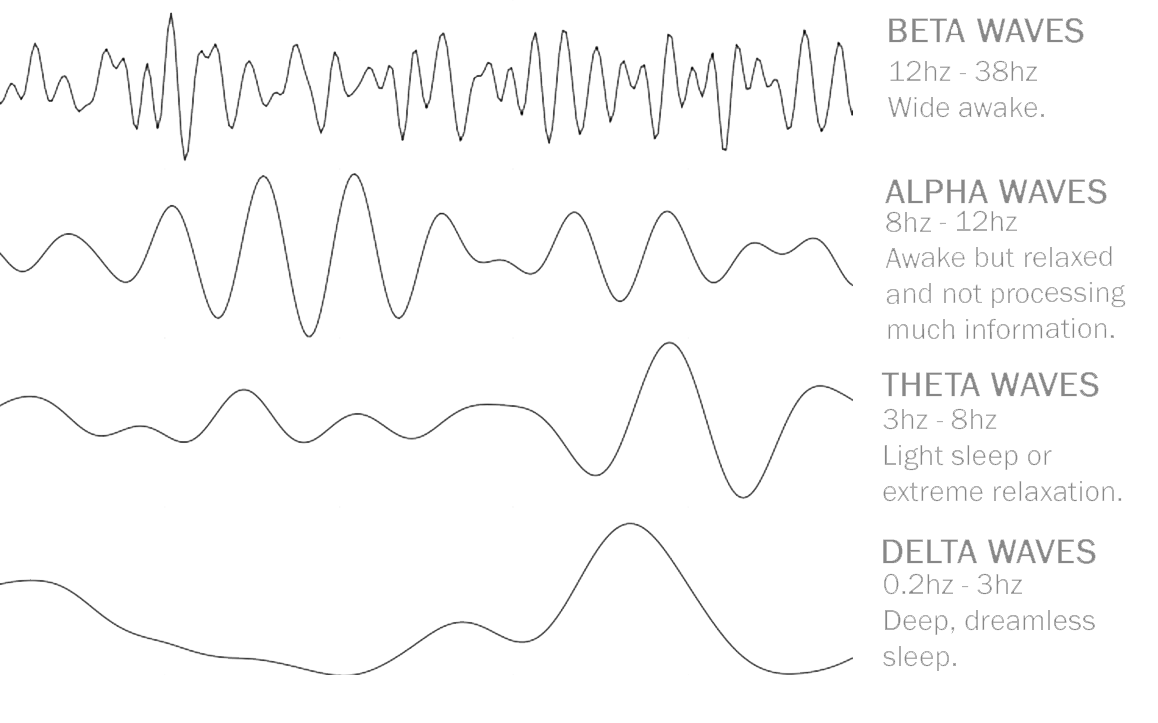
We can divide the activity of our brain into different “waves” defined as oscillations of different intensities and frequencies. Brain waves come in four principal frequencies. Starting with the higher intensity, delta, theta, alpha, and beta, and gamma as the lowest. We can measure these oscillations in cycles per second or hertz (Hz). Beta waves (12 to 38 Hz) are high-frequency and low-amplitude brain waves commonly found in an awake state and related to logical thinking and memory.
Monitoring the activity of the participants’ brain with electroencephalography, Dr. Hanslmayr and colleagues found that the power of the beta waves in the posterior area decreases after the stimulation, a state that is beneficial for stimulus processing and memory.
Other studies showed that magnetic stimulation inhibits brain activity and the prefrontal cortex inhibits the posterior areas of the brain. These results make researchers theorize that memory is enhanced thanks to disinhibition from the prefrontal part of the brain over the posterior region.
Overall, the results highlight the complexity of the network that operates in our brains, showing how different regions can be high-wired, controlling how the data is stored or discarded in each situation.
Although more research is needed, magnetic brain stimulation is a promising treatment for people with dementia or memory problems.
References
- van der Plas, M., Braun, V., Stauch, B. J., & Hanslmayr, S. (2021). Stimulation of the left dorsolateral prefrontal cortex with slow rTMS enhances verbal memory formation. PLoS biology, 19(9), e3001363. https://doi.org/10.1371/journal.pbio.3001363
- Buskila Y, Bellot-Saez A, Morley JW. Generating Brain Waves, the Power of Astrocytes. Front Neurosci. 2019;13:1125. Published 2019 Oct 18. doi:10.3389/fnins.2019.01125.
- Zhang G, Yao L, Zhang H, Long Z, Zhao X. Improved working memory performance through self-regulation of dorsolateral prefrontal cortex activation using real-time fMRI. PLoS One. 2013;8(8):e73735. Published 2013 Aug 27. doi:10.1371/journal.pone.0073735
Illustrated by Sofia Polcownuk


[…] post Magnetic Brain Stimulation Can Boost Our Memory first appeared on United Academics […]